News
How do physical forces spread in neurons?
Published in Neuroscience News.
Researchers provide the most detailed description to date of this process at the molecular level, which is key to understanding how cells regulate their functions during embryo development and touch sensation. The findings are published in Nature Physics.
How do embryos develop? Why does the mammalian’s brain cortex fold? How do we feel touch at our fingertips? These and other fundamental biological questions remain unsolved. Yet, they all rely on a common principle: the conversion of a physical stimulus into a biochemical signal.
The field of biology has recently gained new insights into which physical signals travel across cells and how far they spread. One key finding is that such propagation is deeply influenced by some properties of the cell membrane, such as how it deforms and flows under stress. Still, many details of this intricate mechanism remain unclear.
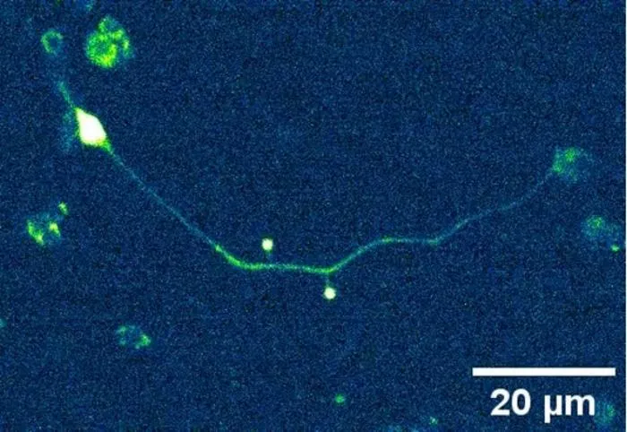
Researchers from ICFO – the Institute of Photonic Sciences (Barcelona, Spain), together with the University of California San Diego, have now shed more light on how neurons transmit strains and stresses through their membranes. In a Nature Physics article, they present the most detailed description to date of the molecular processes underlying this phenomenon by examining isolated neurons of the roundworm Caenorhabditis elegans.
To investigate this, they used an optical tweezer apparatus, a tool based on highly focused laser beams that can both manipulate microscopic objects and measure forces with extraordinary precision. In their experiments, the researchers attached two plastic microspheres to the axons or neurites of the isolated neurons, pulled them with the optical tweezers, and measured how the generated tension traveled from one to the other.
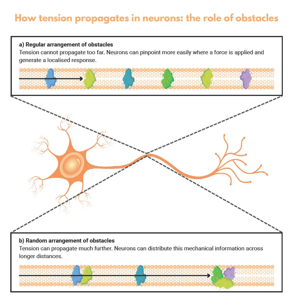
The results showed that tension propagation is faster in touch receptors than in proprioceptors (which sense rapid deformations of the body itself during movement). Even more intriguingly, the researchers found that propagation is influenced not just by the presence of obstacles in the membrane (mainly embedded proteins) but also by how these obstacles are arranged.
Apparently, when they are aligned in a regular pattern, propagation is restricted to shorter distances. According to the researchers, a controlled, limited spread of tension may help neurons pinpoint where a force is applied, distinguish between different stimuli, and generate localized responses without affecting the entire cell. In contrast, a random arrangement of obstacles allows tension to travel much farther, potentially helping cells distribute mechanical information across longer distances. In this way, protein arrangement acts as a regulatory switch: it can keep signals concentrated and localized or let mechanical information travel further through the neuron.
Looking ahead, the researchers plan to explore other interactions of the cell with its environment, many of which have been largely ignored. “It may even be that plasma membrane tension itself regulates obstacles in a feedback/forward loop,” speculates Prof. at ICFO Michael Krieg, lead author of the article.
For now, the study already marks a major advance in mechanobiology. Dr. Eva Kreysing, expert in developmental neuroscience from the University of Cambridge who was not involved in the work, said to the Nature Physics journal: “This is a very timely paper. Given the important part that membrane tension has been shown to play in the regulation of cell function, it is very important to understand how localised this parameter is or how far it propagates.”

Reference:
Català-Castro, F., Bonilla-Quintana, M., Sanfeliu-Cerdán, N. et al. Obstacles regulate membrane tension propagation to enable localized mechanotransduction. Nat. Phys. 21, 1741–1752 (2025).